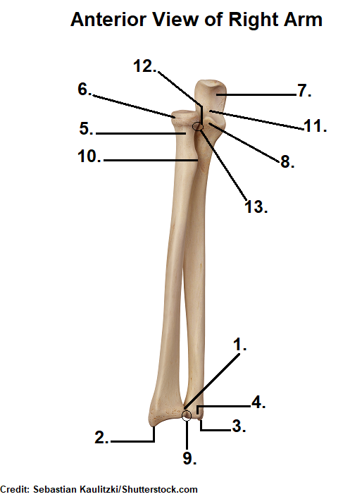
Radius Bone Labelled Diagram - Pivot Joint Definition Examples Function Facts Britannica - I'm not sure of what you mean by bone diagram.. Illustration with skeleton of human hand ulna, radius and humerus bones. I'm not sure of what you mean by bone diagram. This ulnar view labelled illustration is from 'asklepios atlas of the human anatomy'. The ulna articulates with the trochlea and the radius articulates with the capitulum. At the humerus, they articulate with the condyle.
Individually selectable every part, ideal for learning. Proximal radius (head, neck and tuberosity). Labeling activities for anatomy &. 12 photos of the labelled diagram of radius bone. Diagrams at penn foster college.
Femur Definition Function Diagram Facts Britannica from cdn.britannica.com The radius is a long bone in the forearm. Input and provides segmentation of the radius bone in the form. Bones arm hand wrist anatomical anatomy and carpal forearm illustration labels phalanges phalanx radius ray skeleton ulna x. The radius and ulna are the two bones of the forearm. The radius bone is the lateral bone of the forearm and is homologous with the tibia of the lower limb. The bones shown in the chest and hip region in the labeled human skeleton diagram are the ribs humerus is located in the upper arm. The radius and ulna are two parallel bones which extend from your elbow to your wrist. Labeling activities for anatomy &.
Radius bone is a photograph by asklepios medical atlas which was uploaded on august 3rd, 2016.
Each bone is a complex living organ that is made up of many cells, protein fibers, and minerals. The ulna is usually slightly longer than the radius, but the radius is thicker. The radius is considered the most commonly fractured bone in the human body, with distal radius fractures being the most common form of radial. The anatomy of the femur can be divided into proximal, central, distal, and posterior parts. Labeled medical scheme with humerus, muscle, radius and ulna isolated closeup. The ulna articulates with the trochlea and the radius articulates with the capitulum. The skeleton acts as a scaffold by providing support and protection for the soft tissues that make up the rest of the body. Radius, in anatomy, the outer of the two bones of the forearm when viewed with the palm facing forward. Bone markings of the radius & ulna. This ulnar view labelled illustration is from 'asklepios atlas of the human anatomy'. Long bone labeling diagram quizlet from o.quizlet.com. 12 photos of the labelled diagram of radius bone. It is simulated by using a 12 kg/cm servo motor with gears.
The anatomy of the femur can be divided into proximal, central, distal, and posterior parts. The radius and ulna together constitute the forearm. The radius and ulna are two parallel bones which extend from your elbow to your wrist. Each bone is a complex living organ that is made up of many cells, protein fibers, and minerals. (ii) name the structure labelled a, which attaches muscle to bone.
Radius And Ulna Bone Quiz Anatomy from www.registerednursern.com Illustration with skeleton of human hand ulna, radius and humerus bones. Labeled medical scheme with humerus, muscle, radius and ulna isolated closeup. Proceedings of 20th iranian conference on biomedical engineering (icbme 2013). The anatomy of the femur can be divided into proximal, central, distal, and posterior parts. A basic human skeleton is studied in schools with a simple diagram. Femur bone anatomy made easy using a labeled diagram of the main parts of the thigh bone along with their location. The bones shown in the chest and hip region in the labeled human skeleton diagram are the ribs humerus is located in the upper arm. Radius, in anatomy, the outer of the two bones of the forearm when viewed with the palm facing forward.
It's not that clear on this model here, but i'll switch over to another diagram and show you.
12 photos of the labelled diagram of radius bone. General features of a long bone. Glands of the body diagram. Label bone diagram s are getting used for different functions from past many years. Labelled diagram of radius bone. Labeling activities for anatomy &. Left human arm is designed based on original size of relevant human bones. The radius is the bone which is present laterally, which means when your palm is facing upwards, it is away from the middle of your body. Proceedings of 20th iranian conference on biomedical engineering (icbme 2013). The radius and ulna are the two bones of the forearm. Diagram of wrist have bone fracture. (ii) name the structure labelled a, which attaches muscle to bone. The skeleton acts as a scaffold by providing support and protection for the soft tissues that make up the rest of the body.
Glands of the body diagram. Learn everything about the anatomy of radius and ulna with our articles, video tutorials, labeled diagrams, and quizzes. Diagram of wrist have bone fracture. Diagrams at penn foster college. Left human arm is designed based on original size of relevant human bones.
Complete Anatomy Of Radius Bone Learn With Pictures And Quizzes Earth S Lab from www.earthslab.com Radius bone with labels.gif 800 × 800; Left human arm is designed based on original size of relevant human bones. The radius bone is the lateral bone of the forearm and is homologous with the tibia of the lower limb. Labelled diagram of radius bone. I'm not sure of what you mean by bone diagram. The radius is considered the most commonly fractured bone in the human body, with distal radius fractures being the most common form of radial. The radius bone is shorter. Educational labeled scheme with skeleton bone structure description.
The ulna is usually slightly longer than the radius, but the radius is thicker.
As soft tissues like tendons, muscles. Labeling activities for anatomy &. It is simulated by using a 12 kg/cm servo motor with gears. The radius bone is this bone here and it lies laterally in the anatomical position. Healthy body parts example for physiology handout. Labeled medical scheme with humerus, muscle, radius and ulna isolated closeup. The ulna is usually slightly longer than the radius, but the radius is thicker. I'm not sure of what you mean by bone diagram. The radius and ulna are the two bones of the forearm. The bones shown in the chest and hip region in the labeled human skeleton diagram are the ribs people interested in long bone diagram to label also searched for. General features of a long bone. Glands of the body diagram. The skeleton acts as a scaffold by providing support and protection for the soft tissues that make up the rest of the body.
This ulnar view labelled illustration is from 'asklepios atlas of the human anatomy' labelled radius bone. Bone marrow lacks the rigidity of the surrounding bone.


0 Komentar